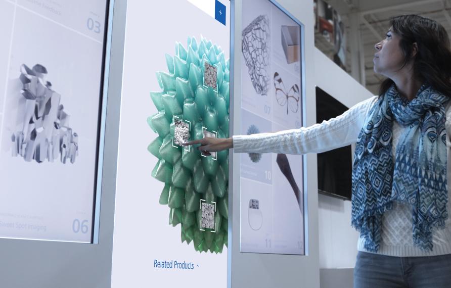


Navigating with Connected Microscopes
With Dr. Sina Shahbazmohamadi


Finding the needle in a haystack.
From the lab of Dr. Sina Shahbazmohamadi
A major challenge in materials characterization is to interrogate microscopic details from a single spot with an unknown location in a comparatively enormous component. This challenge is acute in the world of microelectronics such as multilayer ceramic capacitors (MLCCs). It first requires location, then precise navigation, then observation.
Microscopes excel in different length scales, so finding the needle in a haystack relies on multiple instruments that work well together. Dr. Sina Shahbazmohamadi begins with XRM to establish a “3D roadmap” of devices like MLCCs. This roughly gets to a region of interest. He then moves to the Crossbeam FIB-SEM to target and characterize failures or identify potential counterfeit components.

Needle in a haystack
Quick inspection of the 3D XRM data revealed an anomalous region in one of the MLCC nickel plate layers, which was then targeted for high resolution FIB-SEM analysis.


Virtual view inside the MLCC
X-ray microscopy allows researchers to inspect the inside of the device for voids or defects prior to cutting it open.

Visualizing defects with FIB-SEM
A femtosecond laser was user to access the anomalous region in the MLCC that was seen by X-ray microscopy. The region was then characterized by FIB slicing, SEM imaging and EDS mapping to visualize an enlargement and void spaces in the nickel plates.


“When we don't see what we are looking for, we don't give up. Instead, we REFINE our resolution.”
Equally at home in UCONN’s Material Science and Biomedical engineering departments, Dr. Sina Shahbazmohamadi specializes in correlative microscopy and 3D imaging. Recently he’s applied his diverse skillset to develop new testing methods for electrical components across a variety of manufacturing fields.
Experience more breakthroughs and download Dr. Sina Shahbazmohamadi’s full research report.

Connect with us to go deeper on this research and receive updates on future breakthroughs.

Join us live
Locate a ZEISS event near you and discover the power of possibilities.